


Ankle arthroscopy
Arthroscopy of the ankle
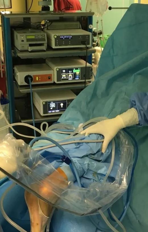
Arthroscopy is a minimally invasive surgery used to address ankle problems.
This technique employs a fibre-optic camera to magnify images inside the joints, aiming to diagnose and treat various ankle and foot disorders without opening the joint itself.
Arthroscopic surgery can be used to treat or diagnose a wide range of foot and ankle conditions, including:
Ankle fractures
Ankle arthroscopy can be used alone or, more commonly, in conjunction with open fracture repair techniques. This can help restore normal alignment of bones and cartilage. It can also be used during tibia or astragalus fracture repairs to assess cartilage injuries inside the ankle.
Ankle instability
Ankle ligaments are structures responsible for maintaining ankle stability. A partial or complete rupture of these ligaments can lead to instability, causing a feeling of giving way. These ligaments are commonly repaired with open surgery. In recent years, arthroscopic techniques have been developed and can be used with appropriate indications to address this problem.
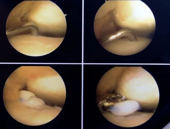
Anterior ankle impingement
This problem occurs when bone or soft tissues in the front of the ankle impede normal joint movement during ankle flexion and extension. Symptoms include ankle pain and swelling. More commonly, this condition limits the ability to dorsiflex the ankle. Walking uphill can be painful. Any bony spurs (osteophytes) can be identified with X-rays or magnetic resonance imaging (MRI). In this case, arthroscopy is the best method for removing osteophytes and inflamed soft tissues.
Posterior ankle impingement
This occurs when bone and soft tissues on the back of the ankle become inflamed due to repeated stress. It often causes limited plantar flexion of the ankle, pain, and swelling. This overload syndrome occurs more frequently in dancers but can be observed in other athletes. It may be associated with an accessory bone called os trigonum. In this case, arthroscopy via the posterior approach is the best method for resolving the problem.
Arthrofibrosis
Following trauma or repeated microtrauma to the ankle, excessive scar tissue can form within the ankle. This can lead to a painful and stiff joint condition known as arthrofibrosis. Ankle arthroscopy can be used to identify and remove the scar tissue.
Infection
Infection in the joint space cannot be treated solely with antibiotics. Urgent surgical intervention is often required to clean the joint, and surgical cleaning in arthroscopy provides excellent joint visibility for the removal of necrotic tissue. With the introduction of continuous water, effective washing is facilitated for bacteria removal.
Synovitis
The lining of the soft tissues of the ankle joint (synovial tissue) can become inflamed, leading to pain, swelling, and loss of movement. This can be caused by acute trauma, inflammatory conditions (e.g., rheumatoid arthritis), overuse, or degenerative joint disease (osteoarthritis). In this case, ankle arthroscopy can be used to surgically remove the inflamed tissue that does not respond to non-surgical treatment.
Intrarticular loose bodies
Cartilage, bones, and scar tissue can become free and float within the joint, forming what is known as an intrarticular loose body. Loose bodies can be painful and can cause problems such as locking and cracking. Ankle arthroscopy can be used to locate and remove loose bodies.
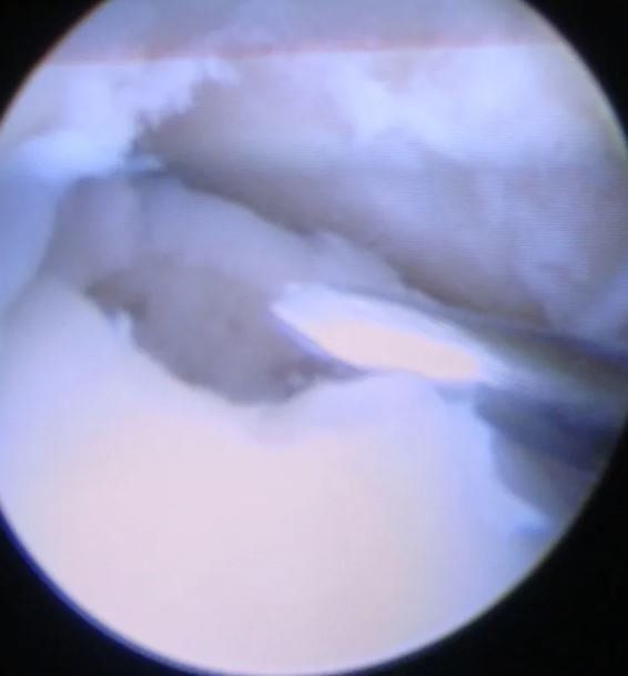
Cartilaginous defect or osteochondral defect
These are localised areas with damaged cartilage and subchondral bone (the bony portion beneath the cartilage) in the ankle joint. Osteochondral defects are usually caused by ankle injuries, such as fractures and sprains. Common symptoms include ankle pain and swelling. Patients often complain of locking and cracking in the ankle. Diagnosis is made using X-rays and magnetic resonance imaging (MRI). CT scans can be used to better evaluate any injuries that extend into the bony portion. Treatment depends on the size, location, and stability of the osteochondral defect. Typically, surgery involves removing the damaged cartilage and creating microperforations in the bone at the site of the lesion to promote healing. In recent years, cartilage substrates and artificial scaffolds have been developed to allow for cartilage recolonisation at the site of the lesion after decontamination.
Ankle arthritis
Ankle fusion is an appropriate treatment option for many patients with end-stage arthritis. Ankle arthroscopy offers a minimally invasive way to perform fusion. Results can be even better than with open techniques, provided the ankle is aligned and there are no concomitant deformities.
Who is not a candidate for arthroscopy?
Elective arthroscopy is not suitable for some patients. Patients with severe arthritic changes with loss of joint space, associated with severe deviation, may not benefit from arthroscopic fusion procedures. Active infections or other health problems may compromise the possibility of undergoing arthroscopy.
Possible complications
Complications are those that can occur in any surgical procedure, although arthroscopy usually has a lower incidence of these complications. These include anaesthetic complications, infections, and bleeding.
Potential specific complications of ankle arthroscopy include injuries to nerves, blood vessels, tendons, ligaments, or cartilage. Numbness, weakness, or tingling in the foot due to transient neurological damage may occur in about 10 per cent of cases and usually resolves over time.
Frequently Asked Questions (FAQs)
When can I return to driving?
You can return to driving when you are able to bear weight without any limitation and are no longer taking pain medication. This generally takes from 2 weeks to two months depending on the cases.
When can I expect to return to work or sports?
For jobs that do not involve heavy physical exertion (e.g., office work), you may be able to return to work a few days after surgery. Most patients should expect to be away from work for at least one or two weeks.
As for sports, it is possible to resume high-level activity following a recovery period of at least 4-6 weeks.
What are the results of arthroscopy?
With the right indication for arthroscopic treatment and proper technical execution, 70-90% of patients undergoing arthroscopy for common issues achieve good or excellent results.
What are the advantages of arthroscopy?
Arthroscopy enables direct visualisation of the inside of the ankle without requiring large scars. This minimises problems associated with large incisions in conventional surgery, such as infections and pain. The minimally invasive nature of this procedure involves a very short hospital stay, typically 24-48 hours. Patients may be able to start functional rehabilitation more quickly and, consequently, return to work and sports much sooner.


